文献:Hydrophobic insertion-based engineering of tumor cell-derived exosomes for SPECT/NIRF imaging of colon cancer
文献链接:https://link.springer.com/article/10.1186/s12951-020-00746-8
作者:Boping Jing, Yongkang Gai, Ruijie Qian, Zhen Liu, Ziyang Zhu, Yu Gao, Xiaoli Lan and Rui An
相关产品:
DSPE-PEG2000-FITC(磷脂-聚乙二醇-荧光素)
DSPE-PEG2000-Cy5(磷脂-聚乙二醇-花菁染料CY5 )
DSPE-PEG2000-HYNIC(磷脂-聚乙二醇-HYNIC)
DSPE-PEG2000-Cy7(磷脂-聚乙二醇-花菁染料Cy7)
原文摘要:
Background: Tumor cell-derived exosomes (TEx) have emerged as promising nanocarriers for drug delivery. Noninvasive multimodality imaging for tracing the in vivo trafcking of TEx may accelerate their clinical translation. In this study, we developed a TEx-based nanoprobe via hydrophobic insertion mechanism and evaluated its performance in dual single-photon emission computed tomography (SPECT) and near-infrared fuorescence (NIRF) imaging of colon cancer.Results: TEx were successfully isolated from HCT116 supernatants, and their membrane vesicle structure was confrmed by TEM. The average hydrodynamic diameter and zeta potential of TEx were 110.87±4.61 nm and –9.20±0.41 mV, respectively. Confocal microscopy and fow cytometry fndings confrmed the high tumor binding ability of TEx. The uptake rate of 99mTc-TEx-Cy7 by HCT116 cells increased over time, reaching 14.07±1.31% at 6 h of co-incubation. NIRF and SPECT imaging indicated that the most appropriate imaging time was 18 h after the injection of 99mTc-TEx-Cy7 when the tumor uptake (1.46%±0.06% ID/g) and tumor-to-muscle ratio (8.22±0.65) peaked. Compared with radiolabeled adipose stem cell derived exosomes (99mTc-AEx-Cy7), 99mTc-TEx-Cy7 exhibited a signifcantly higher tumor accumulation in tumor-bearing mice.Conclusion: Hydrophobic insertion-based engineering of TEx may represent a promising approach to develop and label exosomes for use as nanoprobes in dual SPECT/NIRF imaging. Our fndings confrmed that TEx has a higher tumor-targeting ability than AEx and highlight the potential usefulness of exosomes in biomedical applications
DSPE是一种磷脂。它有一个亲水性的头部(磷酸乙醇胺部分)和两条疏水性的长链脂肪酸(硬脂酸)尾部。PEG2000(聚乙二醇,分子量为 2000)是一个亲水性的聚合物链。它通过共价键连接到 DSPE 的头部。这种结构使得分子一端是亲脂性的,另一端是亲水性的,呈现出两亲性的特点。DSPE - PEG2000 在水中具有良好的溶解性。同时,因为 DSPE 部分的存在,它也能够在一些有机溶剂中溶解,特别是那些既能与亲脂性成分又能与亲水性成分相互作用的溶剂。该文献基于此类产品的特性在成像方面有应用,制备如下:

图:FITC结构式
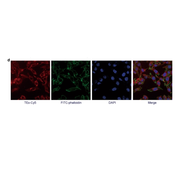
采用荧光细胞术分析TEx的荧光强度,以确定其内化水平。以一定的剂量孵育DSPE-PEG2000-FITC在室温下放置,FITC-TEx通过离心过滤装置。为内化实验,HCT116细胞接种于不同浓度的FITC-TEx处理。孵化后发酵,细胞消化溶解于 PBS用于流式细胞术分析。通过共聚焦显微镜观察TEx的tumour结合能力。TEx和DSPE-PEG2000-Cy5在室温下孵育,得到cy5标记的TEx (TEx- cy5)。将TEx-Cy5 加入HCT116细胞在共聚焦皿中培养,并孵育不同时间用4,6-Diamidino-2-phenylindole (DAPI)反染。细胞固定在荧光显微镜下观察。将10个TEx-Cy5加入到共聚焦培养皿中培养的HCT116细胞中孵育。tumour细胞的细胞骨架用FITC-吞噬素染色,细胞核用DAPI染色。用多聚甲醛加热细胞,在共聚焦显微镜下观察。
一定剂量的DSPE-PEG2000-HYNIC和一定剂量的DSPE-PEG2000-Cy7与TEx或AEx 在室温下形成HYNIC(Cy7)- PEG2000-DSPE-TEx(HYNIC-TEx-Cy7)和HYNIC(Cy7)-PEG2000-DSPE-AEx(HYNIC-AEx-Cy7),如前所述。在进一步使用前,Te样品通过离心烧瓶装置。

图:荧光标记图像
结论:
该文献制备的纳米探针被成功地用于多模态/NIRF成像。疏水相互作用的利用为工程基于外泌体的多模态显像剂提供了可能。证实了DSPE-PEG2000功能化组可以使用这种方法插入到外泌体的表面。该研究还证明,tumour细胞外泌体是一种潜在的高质量的多模态成像纳米载体,具有应用前景。

 2024-12-18 作者:ws 来源:

